• P-ISSN 0974-6846 E-ISSN 0974-5645

Indian Journal of Science and Technology

Article

Indian Journal of Science and Technology

Year: 2024, Volume: 17, Issue: 13, Pages: 1368-1380

Original Article

A Novel 3D Multi-Layer Convolutional Neural Networks for Lung Cancer Segmentation in CT Images

Received Date:16 August 2023, Accepted Date:27 February 2024, Published Date:28 March 2024

Abstract

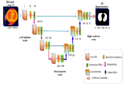
Background/Objectives: A novel three-dimensional efficient Multi-Layer Convolutional Neural Network (3D-MLCNN) is proposed for detecting lung tumors accurately using Computerized Tomography (CT) lung tumor images. The proposed K-means segmentation algorithm for labeling the tumor region automatically. This proposed K-means segmentation algorithm automatically labels the tumor regions to process with the 3D MLCNN model to predict tiny tumors and extract tumor regions accurately. Methods: The proposed 3DMLCNN network goal is to extract the tumor region in CT lung images to classify the lung tumor volume by pixel-wise segmentation model. Findings: The proposed 3D MLCNN segmentation model for detecting the segmenting tumors produces outperforming results for predicting even tiny tumors in the lung images. Experimental results demonstrated with lung cancer CT images in TCIA datasets show that the proposed model 3D-MLCNN achieved a dice coefficient (9.6%), Intersection over Union (IoU) (80%), F1-Score (9.33%), Sensitivity (17.11%), and Accuracy (98%) respectively. However, the proposed model 3D MLCNN was evaluated and compared with the existing state-of-the-art segmentation methods, which shows a 10% improvement in the segmentation process. Novelty: A novel 3D MLCNN model enhances the tumor region and predicts the tumor accurately by labeling the tumor using K-means labeling techniques.

Keywords: 3D Convolutional Neural Networks, Lung Cancer CT Images, K - Means Labeling, Feature Visualization, Deep Learning

References

  1. Yang J, Wu B, Li L, Cao P, Zaiane O. MSDS-UNet: A multi-scale deeply supervised 3D U-Net for automatic segmentation of lung tumor in CT. Computerized Medical Imaging and Graphics. 2021;92:101957. Available from: https://doi.org/10.1016/j.compmedimag.2021.101957
  2. Dou Q, Yu L, Chen H, Jin Y, Yang X, Qin J, et al. 3D deeply supervised network for automated segmentation of volumetric medical images. Medical Image Analysis. 2017;41:40–54. Available from: https://doi.org/10.1016/j.media.2017.05.001
  3. Zhao D, Liu Y, Yin H, Wang Z. An attentive and adaptive 3D CNN for automatic pulmonary nodule detection in CT image. Expert Systems with Applications. 2023;211:118672. Available from: https://doi.org/10.1016/j.eswa.2022.118672
  4. Gan W, Wang H, Gu H, Duan Y, Shao Y, Chen H, et al. Automatic segmentation of lung tumors on CT images based on a 2D & 3D hybrid convolutional neural network. The British Journal of Radiology. 2021;94(1126):1–9. Available from: https://doi.org/10.1259/bjr.20210038
  5. Nishio M, Muramatsu C, Noguchi S, Nakai H, Fujimoto K, Sakamoto R, et al. Attribute-guided image generation of three-dimensional computed tomography images of lung nodules using a generative adversarial network. Computers in Biology and Medicine. 2020;126:104032. Available from: https://doi.org/10.1016/j.compbiomed.2020.104032
  6. Song E, Long J, Ma G, Liu H, Hung CC, Jin R, et al. Prostate lesion segmentation based on a 3D end-to-end convolution neural network with deep multi-scale attention. Magnetic Resonance Imaging. 2023;99:98–109. Available from: https://doi.org/10.1016/j.mri.2023.01.015
  7. Cifci MA. SegChaNet: A Novel Model for Lung Cancer Segmentation in CT Scans. Applied Bionics and Biomechanics. 2022;2022:1–16. Available from: https://doi.org/10.1155/2022/1139587
  8. Najeeb S, Bhuiyan MIH. Spatial feature fusion in 3D convolutional autoencoders for lung tumor segmentation from 3D CT images. Biomedical Signal Processing and Control. 2022;78:103996. Available from: https://doi.org/10.1016/j.bspc.2022.103996
  9. Apostolopoulos ID, Papathanasiou ND, Panayiotakis GS, Panayiotakis. Classification of lung nodule malignancy in computed tomography imaging utilizing generative adversarial networks and semi-supervised transfer learning. Biocybernetics and Biomedical Engineering. 2021;41(4):1243–1257. Available from: https://doi.org/10.1016/j.bbe.2021.08.006
  10. Xu R, Wang C, Xu S, Meng W, Zhang X. Dual-stream Representation Fusion Learning for accurate medical image segmentation. Engineering Applications of Artificial Intelligence. 2023;123(Part B):106402. Available from: https://doi.org/10.1016/j.engappai.2023.106402
  11. Zhao J, Dang M, Chen Z, Wan L. DSU-Net: Distraction-Sensitive U-Net for 3D lung tumor segmentation. Engineering Applications of Artificial Intelligence. 2022;109:104649. Available from: https://doi.org/10.1016/j.engappai.2021.104649
  12. Li L, Zhao X, Lu W, Tan S. Deep learning for variational multimodality tumor segmentation in PET/CT. Neurocomputing. 2020;392:277–295. Available from: https://doi.org/10.1016/j.neucom.2018.10.099
  13. MYA, Yang Y, Meher PK, Dakua SP. Dense-PSP-UNet: A neural network for fast inference liver ultrasound segmentation. Computers in Biology and Medicine. 2023;153:1–11. Available from: https://doi.org/10.1016/j.compbiomed.2022.106478
  14. Chi J, Li Z, Sun Z, Yu X, Wang H. Hybrid transformer UNet for thyroid segmentation from ultrasound scans. Computers in Biology and Medicine. 2023;153:106453. Available from: https://doi.org/10.1016/j.compbiomed.2022.106453
  15. Li B, Keikhosravi A, Loeffler AG, Eliceiri KW. Single image super-resolution for whole slide image using convolutional neural networks and self-supervised color normalization. Medical Image Analysis. 2021;68:101938. Available from: https://doi.org/10.1016/j.media.2020.101938
  16. Li P, Ma W. OverSegNet: A convolutional encoder–decoder network for image over-segmentation. Computers and Electrical Engineering. 2023;107:108610. Available from: https://doi.org/10.1016/j.compeleceng.2023.108610
  17. Zunair H, Hamza AB. Sharp U-Net: Depthwise convolutional network for biomedical image segmentation. Computers in Biology and Medicine. 2021;136:104699. Available from: https://doi.org/10.1016/j.compbiomed.2021.104699
  18. Lembhe A, Motarwar P, Patil R, Elias S. Enhancement in Skin Cancer Detection using Image Super Resolution and Convolutional Neural Network. Procedia Computer Science. 2023;218:164–173. Available from: https://doi.org/10.1016/j.procs.2022.12.412
  19. Afshar P, Oikonomou A, Naderkhani F, Tyrrell PN, Plataniotis KN, Farahani K, et al. 3D-MCN: A 3D Multi-scale Capsule Network for Lung Nodule Malignancy Prediction. Scientific Reports. 2020;10(1):1–11. Available from: https://doi.org/10.1038/s41598-020-64824-5
  20. Borlea ID, Precup RE, Borlea AB, Iercan D. A Unified Form of Fuzzy C-Means and K-Means algorithms and its Partitional Implementation. Knowledge-Based Systems. 2021;214:106731. Available from: https://doi.org/10.1016/j.knosys.2020.106731
  21. Dheepa G, Chithra PL. An Efficient Encoder-Decoder CNN for Brain Tumor Segmentation in MRI Images. IETE Journal of Research. 2022;p. 1–12. Available from: https://doi.org/10.1080/03772063.2022.2098182
  22. Chithra PL, Dheepa G. Di‐phase midway convolution and deconvolution network for brain tumor segmentation in MRI images. International Journal of Imaging Systems and Technology. 2020;30(3):674–686. Available from: https://doi.org/10.1002/ima.22407
  23. Ansari MY, Yang Y, Balakrishnan S, Abinahed J, Al-Ansari A, Warfa M, et al. A lightweight neural network with multiscale feature enhancement for liver CT segmentation. Scientific Reports. 2022;12(1):1–12. Available from: https://doi.org/10.1038/s41598-022-16828-6
  24. Dakua SP, Abi-Nahed J. Patient-oriented graph-based image segmentation. Biomedical Signal Processing and Control. 2013;8(3):325–332. Available from: https://doi.org/10.1016/j.bspc.2012.11.009
  25. Dakua SP. Use of chaos concept in medical image segmentation. Computer Methods in Biomechanics and Biomedical Engineering: Imaging & Visualization. 2013;1(1):28–36. Available from: https://doi.org/10.1080/21681163.2013.765709

Copyright

© 2024 Chithra & Bhavani. This is an open-access article distributed under the terms of the Creative Commons Attribution License, which permits unrestricted use, distribution, and reproduction in any medium, provided the original author and source are credited. Published By Indian Society for Education and Environment (iSee)

DON'T MISS OUT!

Subscribe now for latest articles and news.