Indian Journal of Science and Technology
Year: 2022, Volume: 15, Issue: 3, Pages: 97-106
Original Article
R Preetha1*, S Vanaja2, K Durga Devi1, R Lathamanju3, Nami Susan Kurian4, Jacqulin Veda Jancy S1
1Assistant Professor (SG), Department of Electronics and Communication Engineering, SRM Institute of Science & Technology
2Assistant Professor (SG), Department of Electronics and Communication Engineering,
Easwari Engineering College, Chennai
3Associate Professor, Department of Electronics and Communication Engineering, SRM
Institute of Science & Technology
4Assistant Professor, Department of Electronics and Communication Engineering,
Rajalakshmi Institute of Technology
*Corresponding Author
Email: [email protected]
Received Date:19 October 2021, Accepted Date:12 December 2021, Published Date:31 January 2022
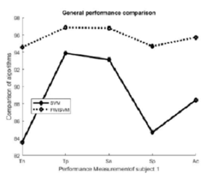
Objective: The objective of the research work is to reconstruct the brain tumor three-dimensionally with high degree of accuracy. Methods: This study describes 3D reconstruction of brain tumor using Mass Sphering Approach (MSA) algorithm. 39 weighted features are extracted from the non-tumor and tumor pixels. These weighted features are used to train the Support Vector Machine (SVM) algorithm. Number of training samples taken to train SVM algorithm are 268 and the testing sample are 64. The complete MR image set of a subject (64 axial slices) are detected for tumor pixels and these slices are concatenated to get volumetric tumor information. Findings: 5-step MSA algorithm is proposed which includes filtering, segmentation, classification, optimization and reconstruction. MR images are subjected to Rician noise which can be removed by a simple correction scheme, initiated to change the bias due to the Rician distribution of the noisy magnitude data. The filtered MR image slices are segmented and classified to detect the tumor areas and the tumor pixels are subjected for 3D reconstruction. The improvement in performance of MSA is depicted by comparing the algorithm with traditional SVM. Novelty: The accuracy achieved in detecting glioma and glioblastoma using MSA are 95.24% and 99% respectively which is highly remarkable.
Keywords: Glioma; voxel; Magnetic Resonance; Classification; Immune; Reconstruction
© 2022 Preetha et al. This is an open-access article distributed under the terms of the Creative Commons Attribution License, which permits unrestricted use, distribution, and reproduction in any medium, provided the original author and source are credited.
Published By Indian Society for Education and Environment (iSee)
Subscribe now for latest articles and news.