• P-ISSN 0974-6846 E-ISSN 0974-5645

Indian Journal of Science and Technology

Article

Indian Journal of Science and Technology

Year: 2023, Volume: 16, Issue: 43, Pages: 3905-3910

Original Article

Visual Acuity: An Eye Parameter to Predict Craniofacial Skeletal Malocclusion

Received Date:05 September 2023, Accepted Date:03 October 2023, Published Date:17 November 2023

Abstract

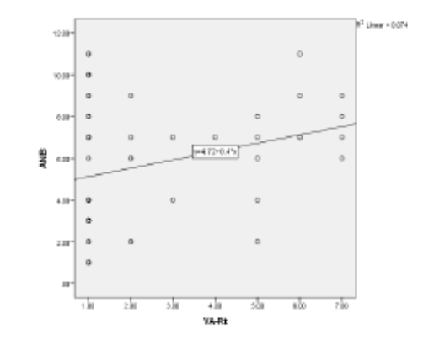
Objectives: The study deals with the identifying correlations between visual acuity and Class I vs Class II skeletal pattern. Methods: A total of seventy subjects were enrolled in the study with age ranging from 11-29 years. Using ANB angle, they were equally divided in two groups (Class I, Class II). Visual acuity of each subject were measured and correlated with each group. Descriptive statistics was compiled and comparison between the groups was made using t-test and Mann-Whitney U test followed by Pearson’s correlation. Level of statistical significance was set at p≤0.05. Findings: The mean age difference between Class I and Class II samples was -0.74 and not statistically significant, indicating that the samples for both categories were of comparable ages. On comparison of visual acuity of right eye, there were 22.7% subjects with myopia in Class I compared to 77.3% in Class II, and this difference in proportion was statistically significant (ES=0.429). In left eye, 25.0% of Class I subjects had myopia, whereas 75.0 % in Class II, and this difference in proportion was also statistically significant (ES=0.328). Positive Pearson’s correlation was found between ANB and Visual acuity of right eye (r=0.165). Novelty: This study concludes that Class II skeletal pattern subjects have more tendency to develop myopia. Further research involving other eye parameters are required to understand the cause-effect relationship.

Keywords: Eye, Visual Acuity, Malocclusion, Opthalmology, Stomatognathic system

References

  1. Niibe K, Zhang M, Nakazawa K, Morikawa S, Nakagawa T, Matsuzaki Y, et al. The potential of enriched mesenchymal stem cells with neural crest cell phenotypes as a cell source for regenerative dentistry. Japanese Dental Science Review. 2017;53(2):25–33. Available from: https://doi.org/10.1016/j.jdsr.2016.09.001
  2. Douarin NML, Dupin E. The “beginnings” of the neural crest. Developmental Biology. 2018;444(Supplement 1):S3–S13. Available from: https://doi.org/10.1016/j.ydbio.2018.07.019
  3. Ozturk T, Ozsaygili C, Topsakal U. Relationship of skeletal malocclusion with eye and hair color in Turkish adolescent patients. APOS Trends in Orthodontics. 2021;11(2):148–155. Available from: https://doi.org/10.25259/APOS_189_2020
  4. Grocott T, Lozano-Velasco E, Mok GF, Münsterberg AE. The Pax6 master control gene initiates spontaneous retinal development via a self-organising Turing network. Development. 2020;147(24):1–13. Available from: https://doi.org/10.1242/dev.185827
  5. Cunha L, Arno D, Corton G, Moosajee M, M. The Spectrum of PAX6 Mutations and Genotype-Phenotype Correlations in the Eye. Genes . 2019;10(12):1–22. Available from: https://doi:10.3390/genes10121050
  6. Lei H, Liu H, Ding Y, Ge L. Immunohistochemical localization of Pax6 in the developing tooth germ of mice. Journal of Molecular Histology. 2014;45(4):373–379. Available from: https://doi.org/10.1007/s10735-014-9564-5
  7. Zieliński G, Filipiak Z, Ginszt M, Matysik-Woźniak A, Rejdak R, Gawda P. The Organ of Vision and the Stomatognathic System—Review of Association Studies and Evidence-Based Discussion. Brain Sciences. 2021;12(1):1–15. Available from: https://doi.org/10.3390/brainsci12010014
  8. Zieliński G, Matysik-Woźniak A, Pankowska A, Pietura R, Rejdak R, Jonak K. High Myopia and Thickness of Extraocular and Masticatory Muscles—7T MRI, Preliminary Study. Journal of Clinical Medicine. 2023;12(12):1–9. Available from: https://doi.org/10.3390/jcm12124166
  9. Zieliński G, Wójcicki M, Rapa M, Matysik-Woźniak A, Baszczowski M, Ginszt M, et al. Masticatory Muscle Thickness and Activity Correlates to Eyeball Length, Intraocular Pressure, Retinal and Choroidal Thickness in Healthy Women versus Women with Myopia. Journal of Personalized Medicine. 2022;12(4):1–15. Available from: https://doi.org/10.3390/jpm12040626
  10. Monaco A, Sgolastra F, Cattaneo R, Petrucci A, Marci MC, D'Andrea PD, et al. Prevalence of myopia in a population with malocclusions. European Journal of Paediatric Dentistry. 2012;13(3 Suppl):256–258. Available from: https://pubmed.ncbi.nlm.nih.gov/23046255/
  11. Bollero P, MRR, Lagana G, Fusco GD, Lione R, Cozza P. Correlations between dental malocclusions, ocular motility, and convergence disorders: a cross-sectional study in growing subjects. Oral Implantology. 2017;10(3):289–294. Available from: https://pubmed.ncbi.nlm.nih.gov/29285332/
  12. Monaco A, Spadaro A, Sgolastra F, Petrucci A, D'Andrea PD, Gatto R, et al. Prevalence of astigmatism in a paediatric population with malocclusions. European Journal of Paediatric Dentistry. 2011;12(2):91–94. Available from: https://pubmed.ncbi.nlm.nih.gov/21668278/
  13. Monaco A, Spadaro A, Sgolastra F, Petrucci A, D'Andrea PD, Gatto R. Prevalence of hyperopia and strabismus in a paediatric population with malocclusions. European Journal of Paediatric Dentistry. 2011;12(4):272–274. Available from: https://pubmed.ncbi.nlm.nih.gov/22185255/
  14. Gupta N, Gupta G, Gupta S, Goyal M. Relationship between Human Eye and Different Divergence of Skeletal Class I Pattern: A Correlative Study. Journal of Mahatma Gandhi University of Medical Sciences and Technology. 2018;3(2):54–60. Available from: https://www.jmgumst.com/doi/JMGUMST/pdf/10.5005/jp-journals-10057-0073
  15. Caruso S, Gatto R, Capogreco M, Nota A. Association of Visual Defects and Occlusal Molar Class in Children. BioMed Research International. 2018;2018:1–4. Available from: https://doi.org/10.1155/2018/7296289
  16. Baldini A, Nota A, Caruso S, Tecco S. Correlations between the Visual Apparatus and Dental Occlusion: A Literature Review. BioMed Research International. 2018;2018:1–12. Available from: https://doi.org/10.1155/2018/2694517
  17. Vompi C, Serritella E, Galluccio G, Pistella S, Segnalini A, Giannelli L, et al. Evaluation of vision in gnathological and orthodontic patients with temporomandibular disorders: A prospective experimental observational cohort study. Journal of International Society of Preventive and Community Dentistry. 2020;10(4):481–490. Available from: https://journals.lww.com/jpcd/fulltext/2020/10040/evaluation_of_vision_in_gnathological_and.15.aspx
  18. Marchili N, Ortu E, Pietropaoli D, Cattaneo R, Monaco A. Dental Occlusion and Ophthalmology: A Literature Review. The Open Dentistry Journal. 2016;10(1):460–468. Available from: https://opendentistryjournal.com/contents/volumes/V10/TODENTJ-10-460/TODENTJ-10-460.pdf
  19. Vitale S, Cotch MF, Sperduto RD. Prevalence of Visual Impairment in the United States. JAMA. 2006;295(18):2158–2163. Available from: https://jamanetwork.com/journals/jama/fullarticle/202836
  20. Singh NK, James RM, Yadav A, Kumar R, Asthana S, Labani S. Prevalence of Myopia and Associated Risk Factors in Schoolchildren in North India. Optometry and Vision Science. 2019;96(3):200–205. Available from: https://journals.lww.com/optvissci/abstract/2019/03000/prevalence_of_myopia_and_associated_risk_factors.8.aspx

Copyright

© 2023 Rastogi et al. This is an open-access article distributed under the terms of the Creative Commons Attribution License, which permits unrestricted use, distribution, and reproduction in any medium, provided the original author and source are credited. Published By Indian Society for Education and Environment (iSee)

DON'T MISS OUT!

Subscribe now for latest articles and news.