• P-ISSN 0974-6846 E-ISSN 0974-5645

Indian Journal of Science and Technology

Article

Indian Journal of Science and Technology

Year: 2024, Volume: 17, Issue: 3, Pages: 286-300

Original Article

Investigating the Effect of the Azo Dye Amaranth on Beta Lactoglobulin by Multi-Spectroscopic Techniques and Molecular Docking: Role in Protein Aggregation

Received Date:29 September 2023, Accepted Date:20 December 2023, Published Date:20 January 2024

Abstract

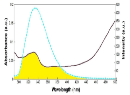
Objectives: This study explored the interaction mechanism between the synthetic azo dye amaranth (AMA) and the widely consumed whey protein beta-lactoglobulin (BLG), using multi-spectroscopic techniques combined with a molecular docking study. Neurodegenerative diseases are often marked by protein aggregation; hence, this study examined the effect of AMA-induced aggregation on BLG at low pH. Methods: The interaction mechanism was investigated at physiological pH using UV–Vis absorption, steady-state and time-resolved fluorescence, Fourier transform infrared (FTIR) spectroscopy, Forster's theory of non-radiation energy transfer, and molecular docking techniques. Biophysical studies, such as turbidity, circular dichroism spectra, and FESEM analysis, have been used to characterize AMA-induced aggregation in BLG. Findings: Steady-state fluorescence quenching of BLG by AMA revealed that the quenching process was dominantly static, owing to complex formation. This was confirmed using time-resolved fluorescence data. BLG showed one binding site for the AMA dye, with a binding affinity of 105 mol/L. According to the FRET analysis, the estimated distance between the binding of BLG and AMA was 3.24 nm. Conformational changes in the BLG were revealed through synchronous fluorescence and FTIR spectroscopy as a result of AMA interaction. Molecular docking studies have suggested that AMA predominantly binds to BLG via hydrogen and hydrophobic bonds. The results of the turbidity experiments showed that AMA concentration affected BLG aggregation. Changes in BLG secondary structure were detected by circular dichroism spectra. FESEM measurements confirmed the amyloid-like structure of the aggregated BLG. Novelty: The experimental results of turbidity analysis revealed that even at low concentrations (0.8 mM) and room temperature, the interaction between the azo dye AMA and BLG can cause protein misfolding, leading to the formation of amyloid aggregates. This research aims to study the harmful effects of edible azo dyes and their ability to initiate the formation of amyloid aggregates linked to various neurodegenerative disorders.

Keywords: Amaranth, Beta­lactoglobulin, Fluorescence quenching, Docking, Amyloid aggregation

References

  1. Alsantali RI, Raja QA, Alzahrani AYA, Sadiq A, Naeem N, Mughal EU, et al. Miscellaneous azo dyes: a comprehensive review on recent advancements in biological and industrial applications. Dyes and Pigments. 2022;199:110050. Available from: https://doi.org/10.1016/j.dyepig.2021.110050
  2. Awuchi CG. Whey Protein from Milk as a Source of Nutraceuticals. In: Egbuna C, Sawicka B, Khan J., eds. Food and agricultural byproducts as important source of valuable nutraceuticals. (pp. 159-183) Springer, Cham. 2022.
  3. Al-Shabib NA, Khan JM, Malik A, Alsenaidy AM, Alsenaidy MA, Husain FM, et al. Negatively charged food additive dye “Allura Red” rapidly induces SDS-soluble amyloid fibril in beta-lactoglobulin protein. International Journal of Biological Macromolecules. 2018;107(Part B):1706–1716. Available from: https://doi.org/10.1016/j.ijbiomac.2017.10.032
  4. Alresaini S, Malik A, Alonazi M, Alhomida A, Khan JM. SDS induces amorphous, amyloid-fibril, and alpha-helical structures in the myoglobin in a concentration-dependent manner. International Journal of Biological Macromolecules. 2023;231:123237. Available from: https://doi.org/10.1016/j.ijbiomac.2023.123237
  5. Warsi MS, Habib S, Talha M, Mir AR, Alam K, Ali A, et al. Characterization of human serum albumin modified by hair dye component, 4-chloro-1,2-phenylenediamine: Role in protein aggregation, redox biology and cytotoxicity. Journal of Molecular Liquids. 2021;331:115731. Available from: https://doi.org/10.1016/j.molliq.2021.115731
  6. Khan MS, Bhatt S, Tabrez S, Rehman MT, Alokail MS, Alajmi MF. Quinoline yellow (food additive) induced conformational changes in lysozyme: a spectroscopic, docking and simulation studies of dye-protein interactions. Preparative Biochemistry & Biotechnology. 2020;50(7):673–681. Available from: https://doi.org/10.1080/10826068.2020.1725774
  7. Khan JM, Malik A, Husain FM, Hakeem MJ, Alhomida AS. Sunset Yellow Dye Induces Amorphous Aggregation in β-Lactoglobulin at Acidic pH: A Multi-Techniques Approach. Polymers. 2022;14(3):1–13. Available from: https://doi.org/10.3390/polym14030395
  8. Chaves OA, Loureiro RJ, Costa-Tuna A, Almeida ZL, Pina J, Brito RM, et al. Interaction of Two Commercial Azobenzene Food Dyes, Amaranth and New Coccine, with Human Serum Albumin: Biophysical Characterization. ACS Food Science & Technology. 2023;3:955–968. Available from: https://doi.org/10.1021/acsfoodscitech.3c00125
  9. Wei Y, Vriesekoop F, Yuan Q, Liang H. β-Lactoglobulin as a Nanotransporter for Glabridin: Exploring the Binding Properties and Bioactivity Influences. ACS Omega. 2018;3(9):12246–12252. Available from: https://doi.org/10.1021/acsomega.8b01576
  10. Salim MM, Sharkasy MEE, Belal F, Walash M. Multi-spectroscopic and molecular docking studies for binding interaction between fluvoxamine and human serum albumin. Spectrochimica Acta Part A: Molecular and Biomolecular Spectroscopy. 2021;252:119495. Available from: https://doi.org/10.1016/j.saa.2021.119495
  11. Xu H, Lu Y, Zhang T, Liu K, Liu L, He Z, et al. Characterization of binding interactions of anthraquinones and bovine β-lactoglobulin. Food Chemistry. 2019;281:28–35. Available from: https://doi.org/10.1016/j.foodchem.2018.12.077
  12. Albani JR, Vogelaer J, Bretesche L, Kmiecik D. Tryptophan 19 residue is the origin of bovine β-lactoglobulin fluorescence. Journal of Pharmaceutical and Biomedical Analysis. 2014;91:144–150. Available from: https://doi.org/10.1016/j.jpba.2013.12.015
  13. Almeida ZL, Brito RMM. Structure and Aggregation Mechanisms in Amyloids. Molecules. 2020;25(5):1–30. Available from: https://doi.org/10.3390/molecules25051195
  14. Al-Shabib NA, Khan JM, Malik A, Rehman MT, Alajmi MF, Husain FM, et al. Molecular interactions of food additive dye quinoline yellow (Qy) with alpha-lactalbumin: Spectroscopic and computational studies. Journal of Molecular Liquids. 2020;311:113215. Available from: https://doi.org/10.1016/j.molliq.2020.113215
  15. Fei S, Zou L, Xie X, Yang F, Chen H, Li X. Purification and Characterization of Bovine β-Lactoglobulin Variants A and B (Characterization of Bovine β-Lactoglobulin Variants) Food Science and Technology Research. 2020;26(3):399–409. Available from: https://doi.org/10.3136/fstr.26.399
  16. Hoppenreijs LJG, Fitzner L, Ruhmlieb T, Heyn TR, Schild K, Goot AJJVD, et al. Engineering amyloid and amyloid-like morphologies of β-lactoglobulin. Food Hydrocolloids. 2022;124(Part A):1–15. Available from: https://doi.org/10.1016/j.foodhyd.2021.107301

Copyright

© 2024 Gayathri & Bakkialakshmi. This is an open-access article distributed under the terms of the Creative Commons Attribution License, which permits unrestricted use, distribution, and reproduction in any medium, provided the original author and source are credited. Published By Indian Society for Education and Environment (iSee)

DON'T MISS OUT!

Subscribe now for latest articles and news.