• P-ISSN 0974-6846 E-ISSN 0974-5645

Indian Journal of Science and Technology

Article

Indian Journal of Science and Technology

Year: 2020, Volume: 13, Issue: 20, Pages: 2030-2040

Original Article

Prediction of different stages in Diabetic retinopathy from retinal fundus images using radial basis function based SVM

Received Date:18 April 2020, Accepted Date:14 May 2020, Published Date:19 June 2020

Abstract

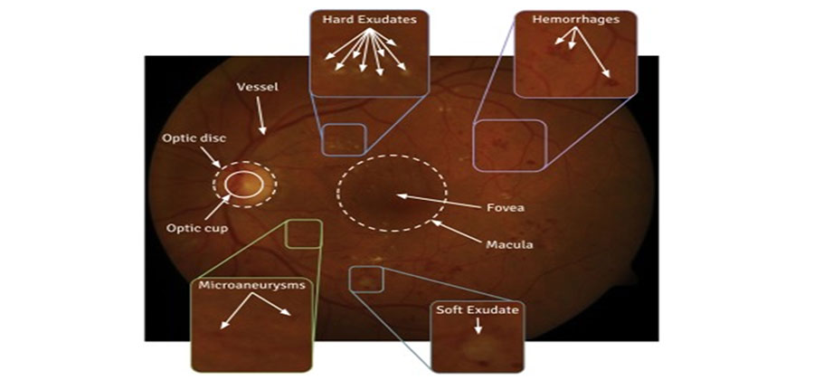
Objectives: This study proposes an automatic computer-aided screening system for prediction of Diabetic retinopathy (DR) by using image processing and machine learning techniques. Method: This proposed model can predict DR in three different stages, Normal, Non-Proliferative Diabetic retinopathy (NPDR) and Proliferative diabetic retinopathy (PDR) based on the features those are present in an input retinal fundus image using support Vector Machine(SVM). For better feature extraction each input retinal fundus image is pre-processed using three techniques; Image compression, Color layer separation and Contrast Limited Adaptive equalization (CLAHE). After pre-processing, the feature extraction is done using different techniques like Linear Spatial filtering, image thresholding and Top-hat operation for extraction of different features like micro aneurysms, blood vessels and exudates respectively. These extracted features are used for designing the classifier. Different kernels of SVM have been applied to the same set of feature and compared. Findings: Finally, Radial Basis Function(RBF) based Kernel SVM outperform others with an accuracy value of 97.2% using a test dataset of size 255 images. Novelty: As the model addresses three class classification of DR with a vast set of feature matrix, it performs well in detection of DR at its earlier state even with minimum feature set.

Keywords: Diabetic retinopathy; Non-proliferative diabetic retinopathy; Proliferative diabetic retinopathy; Support vector machine; Contrast limited adaptive equalization; Radial basis function

References

  1. Khojasteh P, Júnior LAP, Carvalho T, Rezende E, Aliahmad B, Papa JP, et al. Exudate detection in fundus images using deeply-learnable features. Computers in Biology and Medicine. 2019;104:62–69. doi: 10.1016/j.compbiomed.2018.10.031
  2. Medhi JP, Dandapat S. An effective fovea detection and automatic assessment of diabetic maculopathy in color fundus images. Computers in Biology and Medicine. 2016;74:30–44. doi: 10.1016/j.compbiomed.2016.04.007
  3. Naqvi SAG, Zafar MF, Haq Iu. Referral system for hard exudates in eye fundus. Computers in Biology and Medicine. 2015;64:217–235. doi: 10.1016/j.compbiomed.2015.07.003
  4. Youssef D, Solouma NH. Accurate detection of blood vessels improves the detection of exudates in color fundus images. Computer Methods and Programs in Biomedicine. 2012;108:1052–1061. doi: 10.1016/j.cmpb.2012.06.006
  5. Prentašić P, Lončarić S. Detection of exudates in fundus photographs using deep neural networks and anatomical landmark detection fusion. Computer Methods and Programs in Biomedicine. 2016;137:281–292. doi: 10.1016/j.cmpb.2016.09.018
  6. Gupta A, Chhikara R. Diabetic Retinopathy: Present and Past. Procedia Computer Science. 2018;132:1432–1440. doi: 10.1016/j.procs.2018.05.074
  7. Roychowdhury S, Koozekanani DD, Parhi KK. DREAM: Diabetic Retinopathy Analysis Using Machine Learning. IEEE Journal of Biomedical and Health Informatics. 2014;18(5):1717–1728. doi: 10.1109/jbhi.2013.2294635
  8. Pratt H, Coenen F, Broadbent DM, Harding SP, Zheng Y. Convolutional neural networks for diabetic retinopathy. Procedia Computer Science. 2016;90:200–205. doi: 10.1016/j.procs.2016.07.014
  9. Dutta S, Manideep BC, Basha SM, Caytiles RD, Iyengar NCSN. Classification of Diabetic Retinopathy Images by Using Deep Learning Models. International Journal of Grid and Distributed Computing. 2018;11(1):99–106. doi: 10.14257/ijgdc.2018.11.1.09
  10. Argade KS, Deshmukh KA, Narkhede MM, Sonawane NN, Jore S. Automatic detection of diabetic retinopathy using image processing and data mining techniques. 2015 International Conference on Green Computing and Internet of Things (ICGCIoT). 2015. doi: 10.1109/ICGCIoT.2015.7380519
  11. Priya R, Aruna P. Diagnosis of diabetic retinopathy using machine learning techniques. ICTACT Journal on soft computing. 2013;3:563–575.
  12. Antal B, Hajdu A. An Ensemble-Based System for Microaneurysm Detection and Diabetic Retinopathy Grading. IEEE Transactions on Biomedical Engineering. 2012;59(6):1720–1726. doi: 10.1109/tbme.2012.2193126
  13. Gupta A, Issac A, Sengar N, Dutta MK. An efficient automated method for exudates segmentation using image normalization and histogram analysis. Ninth International Conference on Contemporary Computing (IC3). 2016. doi: 10.1109/IC3.2016.7880256
  14. Puranik SS, Malode VB. Morphology based approach for microaneurysm detection from retinal image. 2016 International Conference on Automatic Control and Dynamic Optimization Techniques (ICACDOT). 2016. doi: 10.1109/ICACDOT.2016.7877663
  15. Akkara J, Kuriakose A. Role of artificial intelligence and machine learning in ophthalmology. Kerala Journal of Ophthalmology. 2019;31(2):150. doi: 10.4103/kjo.kjo_54_19

Copyright

© 2020 Behera, Mishra, Ransingh, Chakravarty. This is an open access article distributed under the terms of the Creative Commons Attribution License, which permits unrestricted use, distribution, and reproduction in any medium, provided the original author and source are credited.
Published By Indian Society for Education and Environment (iSee)

DON'T MISS OUT!

Subscribe now for latest articles and news.